
FROM THE STACKS

EDITOR’S NOTE: There are literally thousands of journals published around the world that relate to the disability community. It is virtually impossible to capture even a fraction of them. HELEN receives "stacks" of journals and selectively earmarks what we feel are "must read" articles of interest for our readers. It's a HELEN perk.
ANCOR Issues Findings from 2025 State of America's Direct Support Workforce Crisis Survey

This fall, ANCOR fielded its sixth annual survey of community providers of intellectual and developmental (I/DD) services, continuing the effort to better understand how providers navigate long-term shortages of direct support professionals (DSPs).
This year's survey, which was fielded in August and September, garnered responses from 469 organizations delivering services in 48 states plus the District of Columbia. The findings are outlined in the all-new report, which, since being released this morning, has already gained national news coverage in Axios.
Because Medicaid reimbursement rates have failed to keep pace with inflation and rising costs of living—which has hamstrung community providers’ ability to offer competitive wages—providers are being forced to make impossible choices: either discontinue services for people already supported, or stop accepting new referrals.
As our the report outlines, this puts access to community further from reach at a time when there are already more than 500,000 people with I/DD languishing on states’ waiting lists.
Download the Report (Click here)
The data points are among the key findings from The State of America’s Direct Support Workforce Crisis 2025: The State of America’s Direct Support Workforce Crisis 2025
Caring For Caregivers: How The VA Stipend Sustains Families Of Wounded Warriors
By Haley Fuller
(Military.com - 10/07/25)
Family members who take on the responsibility of caring for a wounded warrior often find themselves balancing financial, emotional, and physical demands that can feel overwhelming. Recognizing this, VA created the Program of Comprehensive Assistance for Family Caregivers (PCAFC). One of its key features is a monthly stipend for primary family caregivers. The purpose is not only to compensate for the time and labor caregiving requires, but also to provide enough support to keep veterans in home or family care rather than in an institutional setting. By offering direct financial aid, VA acknowledges the essential role family members play in the recovery and long-term well-being of disabled veterans.
Eligibility For Veterans And Caregivers
Not every veteran or caregiver qualifies for the stipend, and the criteria are intentionally narrow to ensure the benefit reaches those with the greatest needs. The veteran must have a service-connected disability rating of at least 70%, must have been discharged from the military or be in the process of medically discharing, and must require personal care services on a continuing basis for at least six months. The veteran also must be enrolled in VA health care to access this program.
For caregivers, eligibility depends on being designated as the veteran’s Primary Family Caregiver. This is often parent, spouse, adult child, or another close relative, although in some cases it may be someone living with the veteran full time. Only one primary caregiver may be designated for each veteran, though there can be additional secondary caregivers. The caregiver must complete required training, demonstrate the ability to perform personal care tasks, and commit to providing the needed support on a daily basis.
How The Stipend Is Calculated
The monthly stipend is not a flat rate but varies by location and by the level of care required. VA bases the calculation on the General Schedule Annual Rate for grade 4, step 1 salary for the caregiver’s locality, dividing the annual figure into 12 monthly payments. Veterans assessed as requiring a moderate level of care, sometimes called Level 1, receive 62.5% of this amount. Those determined to be unable to live independently without substantial assistance, or Level 2, receive the full amount.
For example, in Dallas, Texas, a caregiver at Level 1 might receive just over $1,800 per month, while at Level 2 the stipend could exceed $2,900. These figures shift annually as federal pay scales adjust, and they also vary depending on the geographic pay tables established by the Office of Personnel Management.
Additional Benefits Beyond The Stipend
The stipend is the most visible part of the program, but it is not the only form of support. Primary caregivers may qualify for health insurance through CHAMPVA if they do not already have sufficient coverage. They are also eligible for counseling services to address the stress and emotional challenges that come with caregiving. When traveling with a veteran for approved medical care, the caregiver can receive travel reimbursement. Finally, the program offers respite care, giving caregivers temporary relief while ensuring the veteran is safely looked after by another provider.
The Application Process
Applying for the caregiver stipend requires filing VA Form 10-10CG, the official application for PCAFC. This form can be submitted online, by mail, or in person at a VA medical center. Along with the form, applicants must provide documentation showing the veteran’s eligibility, medical needs, and the caregiver’s relationship to the veteran. Training modules and assessments are part of the approval process. Once an application is submitted, the VA conducts evaluations to determine the level of care required and whether the caregiver is prepared to take on the responsibility. Approved caregivers receive monthly payments, and the stipend is treated as non-taxable income.
Challenges In Practice
Although the caregiver stipend program provides much-needed relief, it is not without difficulties. The process of applying can feel familiarly bureaucratic, and delays are common, particularly when medical evidence or training documentation is incomplete. Even once approved, the stipend is subject to reassessment, which means that a change in the veteran’s health or functional ability can raise or lower the amount provided. Many caregivers also emphasize that while the stipend helps, it does not erase the immense physical and emotional toll that comes with round-the-clock care. Because the stipend is classified as compensation rather than wages, it does not carry the same employment protections or benefits that a traditional job might.
Why The Program Matters
For the families of wounded warriors, the caregiver stipend represents more than just financial support. It is a formal acknowledgment by the federal government of the sacrifices families make to keep veterans safe and cared for at home. It also makes possible a higher quality of life for many veterans, who benefit from familiar surroundings and family presence rather than institutional care. The stipend cannot eliminate every burden, but it can make caregiving sustainable and dignified, ensuring those who have already given so much to their country are not left to struggle alone.
Scientists Discover Brain Circuit That Can Switch Off Chronic Pain
J. Nicholas Betley, Nitsan Goldstein
(University of Pennsylvania, Federal Grant sponsored – 10/10/25)
Summary:
Scientists have pinpointed Y1 receptor neurons in the brain that can override chronic pain signals when survival instincts like hunger or fear take precedence. Acting like a neural switchboard, these cells balance pain with other biological needs. The research could pave the way for personalized treatments that target pain at its brain source—offering hope for millions living with long-term pain.

FULL STORY: Fluorescent imaging of NPY+ neurons (green) throughout the brain are shown in addition to neurons in magenta that send projections to the PBN. Credit: J Nicholas Betley
Pain may be unpleasant, but in most cases it plays a vital, even lifesaving, role. Short bursts of pain act as warning signals that protect us from harm. When you touch a hot pan, stub your toe, or bump your head, your nervous system instantly delivers an “Ow!” that prompts you to pull back before more damage occurs. The pain fades, the body heals, and you remember what not to do next time.
Chronic pain, however, is an entirely different story. In this condition, the warning signal doesn’t stop even after the injury has healed. For about 50 million people in the United States, pain becomes a constant, invisible companion that can persist for years or even decades. "It's not just an injury that won't heal," explains neuroscientist at the University of Pennsylvania J. Nicholas Betley, "it's a brain input that's become sensitized and hyperactive, and determining how to quiet that input could lead to better treatments."
Betley, along with collaborators from the University of Pittsburgh and Scripps Research Institute, has discovered an important piece of the chronic pain puzzle. Their research points to a specific group of brainstem cells called Y1 receptor (Y1R)-expressing neurons, located in the lateral parabrachial nucleus (lPBN). These neurons are activated in persistent pain states, but they also process signals related to hunger, fear, and thirst. This suggests that the brain can adjust pain responses when other, more urgent needs demand attention.
The findings, published in Nature, indicate that relief may be possible because, as the researchers write, "there are circuits in the brain that can reduce the activity of neurons that transmit the signal of pain."
Tracking pain in the brain
Working with the Taylor lab at the University of Pittsburgh, Betley’s team used calcium imaging to visualize neuron activity in real time in animal models of both short-term and long-term pain. They observed that Y1R neurons did not simply react to quick bursts of pain; instead, they kept firing steadily during prolonged pain, a phenomenon known as “tonic activity.”
Betley compares this to an engine left running even after you’ve parked the car. The pain signals continue to hum in the background even when physical recovery seems complete. This ongoing neural activity may explain why some people continue to feel pain long after an injury or surgery.
The research originated from an unexpected observation Betley made after joining Penn in 2015: hunger seemed to lessen chronic pain.
"From my own experience, I felt that when you're really hungry you'll do almost anything to get food," he says. "When it came to chronic, lingering pain, hunger seemed to be more powerful than Advil at reducing pain."
That insight inspired further investigation. Former graduate student Nitsan Goldstein found that other critical survival states—such as thirst and fear—can also suppress long-term pain. In collaboration with the Kennedy lab at Scripps, the team showed that the brain’s parabrachial nucleus can filter sensory input to quiet pain when immediate survival takes priority.
"That told us the brain must have a built-in way of prioritizing urgent survival needs over pain, and we wanted to find the neurons responsible for that switch," says Goldstein.
A key part of that switch is neuropeptide Y (NPY), a signaling molecule that helps the brain juggle competing needs. When hunger or fear takes priority, NPY acts on Y1 receptors in the parabrachial nucleus to dampen ongoing pain signals.
"It's like the brain has this built-in override switch," Goldstein explains. "If you're starving or facing a predator, you can't afford to be overwhelmed by lingering pain. Neurons activated by these other threats release NPY, and NPY quiets the pain signal so that other survival needs take precedence."
A scattered signal
The researchers also characterized the molecular and anatomical identity of the Y1R neurons in the lPBN. They found that Y1Rneurons didn't form two tidy anatomical or molecular populations. Instead, these neurons were scattered across many other cell types.
"It's like looking at cars in a parking lot," Betley says. "We expected all the Y1R neurons to be a cluster of yellow cars parked together, but here the Y1R neurons are like yellow paint distributed across red cars, blue cars, and green cars. We don't know exactly why, but we think this mosaic distribution may allow the brain to dampen different kinds of painful inputs across multiple circuits."
Explorations of pain treatment
What excites Betley with this discovery is the further exploration of its potential to "use Y1 neural activity as a biomarker for chronic pain, something drug developers and clinicians have long lacked," he says.
"Right now, patients may go to an orthopedist or a neurologist, and there is no clear injury. But they're still in pain," he says. "What we're showing is that the problem may not be in the nerves at the site of injury, but in the brain circuit itself. If we can target these neurons, that opens up a whole new path for treatment."
This research also suggests that behavioral interventions such as exercise, meditation, and cognitive behavioral therapy may influence how these brain circuits fire, just as hunger and fear did in the lab.
"We've shown that this circuit is flexible, it can be dialed up or down," he says. "So, the future isn't just about designing a pill. It's also about asking how behavior, training, and lifestyle can change the way these neurons encode pain."
J Nicholas Betley is an associate professor in the Department of Biology at the University of Pennsylvania's School of Arts & Sciences.
Nitsan Goldstein was a graduate student in the Betley Lab at Penn Arts & Sciences during this study. He is currently a postdoctoral researcher at the Massachusetts Institute of Technology.
Other authors include Michelle Awh, Lavinia Boccia, Jamie R. E. Carty, Ella Cho, Morgan Kindel, Kayla A. Kruger, Emily Lo, Erin L. Marble, Nicholas K. Smith, Rachael E. Villari, and Albert T. M. Yeung of Penn Arts & Sciences; Niklas Blank and Christoph A. Thaiss of Penn's Perelman School of Medicine; Melissa J. Chee and Yasmina Dumiaty of Carleton University; Rajesh Khanna of University of Florida College of Medicine,; Ann Kennedy and Amadeus Maes of Scripps Research Institute; and Heather N. Allen, Tyler S. Nelson and Bradley K. Taylor of the University of Pittsburg.
This research was supported by the Klingenstein Foundation, the University of Pennsylvania School of Arts and Sciences, the National Institutes of Health (grants F31DK131870, 1P01DK119130, 1R01DK133399, 1R01DK124801, 1R01NS134976, F32NS128392, K00NS124190, F32DK135401, T32DK731442, R61NS126026, R01NS120663, R01NS134976-02, R00MH117264, 1DP1DK140021-01), the National Science Foundation Graduate Research Fellowship Program, the Blavatnik Family Foundation Fellowship, the American Neuromuscular Foundation Development Grant, the American Heart Association (25POST1362884), the Swiss National Science Foundation (206668), the Canadian Institutes of Health Research Project Grant (PJT-175156), the Simons Foundation, a McKnight Foundation Scholar Award, and a Pew Biomedical Scholar Award.
Global Status Report on Neurology
(10/12/2025)

Overview
Neurological conditions are the leading cause of ill health and disability, affecting over 1 in 3 people worldwide. The Global status report on neurology presents the first comprehensive global assessment of the public health response to neurological disorders under the Intersectoral global action plan on epilepsy and other neurological disorders 2022–31 (IGAP).
Drawing on data from 102 WHO Member States representing 71% of the world population, the report sets 2022 baseline values for the 10 global targets of IGAP. It captures the global status across governance and advocacy, financing, service delivery and workforce, access to medicines and technologies, brain health promotion and disease prevention, and research and information systems. The analysis reveals critical gaps and implementation barriers, underscoring the urgent need for bold and coordinated action to achieve IGAP targets by 2031. To accelerate progress, the report offers inclusive, evidence-based and actionable recommendations for policymakers, IGAP partners and the global neurology community – ensuring that people with neurological conditions and their families are at the heart of the response.
--
Alzheimer’s Association Welcomes FDA Clearance of First Blood Test for Use in Primary Care to Rule Out Alzheimer’s-Related Amyloid Pathology
(ALZ.org Email – 10/13/2025)
Key Takeaways
Rules out Alzheimer’s disease pathology and aids in assessing other causes of cognitive decline.
Provides a new accessible tool for timely assessment of cognitive symptoms.
Helps primary care providers prevent unnecessary and invasive testing.
Alzheimer’s disease diagnosis remains a multi-step process.
The U.S. Food and Drug Administration (FDA) has cleared the Elecsys pTau181 plasma test, developed by Roche. This marks the first blood-based biomarker test for Alzheimer’s disease cleared for use in the primary care setting specifically to rule out the presence of amyloid pathology.
“This is another important step toward expanding access to Alzheimer’s disease diagnostic tools,” said Joanne Pike, DrPH, Alzheimer’s Association president and chief executive officer. “At the same time, it is important to understand this test is designed to rule out the presence of amyloid plaques. It is not a test that will give an Alzheimer's disease diagnosis, nor is it a standalone tool for detection.”
According to the company, the Elecsys pTau181 blood test is intended for use in adult patients, aged 55 years and older, presenting with signs or symptoms of cognitive impairment. By providing an efficient, less-invasive method to assess the likelihood of amyloid plaques — a core hallmark of Alzheimer’s disease — this test can help primary care physicians determine which patients do not require Alzheimer’s-related follow-up tests, such as PET scans or cerebrospinal fluid analysis. This can significantly improve the diagnostic process by preventing unnecessary specialized testing for individuals whose cognitive issues stem from non-Alzheimer’s causes.
“By using a ‘rule out’ initial tool in the primary care setting, we can help people who are not living with Alzheimer’s get to the root of their cognitive symptoms faster, while ensuring those who may have the disease are referred for appropriate testing, definitive diagnosis and early treatment,” Pike continued. “This approach also empowers primary care clinicians to guide referrals more effectively, allowing specialists to focus on patients most likely to require advanced evaluation and care.”
The Alzheimer’s Association is committed to ensuring blood tests are used appropriately and effectively across all care settings. Earlier this year, the Association published clinical practice guidelines for the use of blood biomarker tests in specialty care settings, which are continually updated to reflect the evolving scientific landscape. While most data around the use of blood tests are from specialty care settings and clinical trials, we recognize these tests are being used in many settings. The Association urges providers across clinical care settings to utilize guidance on blood biomarker testing in conjunction with clinical judgement to support effective and appropriate use of these much needed tools.
The Association believes this latest development, alongside historic investment in Alzheimer’s and dementia research by the National Institutes of Health (NIH) and other entities, continues to accelerate the pace of scientific discovery, leading to better diagnostic tools and effective disease-modifying treatments.
About the Alzheimer's Association
The Alzheimer’s Association is a worldwide voluntary health organization dedicated to Alzheimer’s care, support and research. Our mission is to lead the way to end Alzheimer's and all other dementia — by accelerating global research, driving risk reduction and early detection, and maximizing quality care and support. Our vision is a world without Alzheimer's and all other dementia®. Visit alz.org or call 800.272.3900.
—
Poem For Premature Babies And Their Families
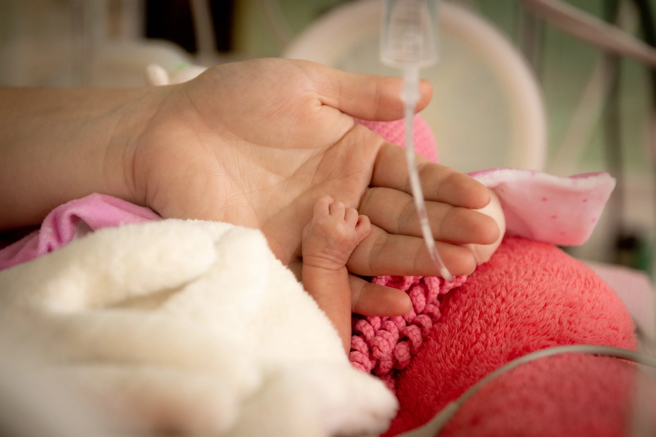
(Written by a preemie Dad Sean Simpson - mumsadvice.co.uk)
Time stands still, the day has arrived
A baby born early, has he survived?
Emotions are torn, what should I do?
Lost in a world that’s so new to you.
The baby is rushed into NICU care
Mum on a table, it doesn’t seem fair
A mother and baby, two floors apart
Separated by distance, connected by heart
You stand on the outside as the scene flashes by
The team go to work, don’t let him die
Tubes and machines keep him alive
You pray for a miracle, let him survive.
The doctors they tell you, prepare for the worst
Your world tears apart your dreams are immersed
He is far too small, he has a battle ahead
You search for some hope in the eyes of the Ped.
The day turns to night and the team goes away
A doctor and nurse are the only to stay
The clock goes so slowly yet the hours still pass
Your try to reach out to that face behind the glass
Your life is the monitor you watch every beat
You watch every breath; you don’t leave your seat
You don’t leave his side he is your blood and your life
You would give your left arm and so would your wife.
A father’s pain is hard yet it only seems token
Compared to a Mum who lays battered and broken
Her dream has been robbed and so has her joy
Her only life’s wish is to hold her new boy.
As night starts to deepen you start to reflect
You start to question what to expect
You try to reason you try to make sense
You feel joy yet your pain is intense
The longest night in history draws to a close
The day that stood still, the day where time froze
The smallest human you have ever seen
So tough, so hard, courage unseen.
Hours become days and days become weeks
Weeks become months, with falls and peaks
The road is long the journey is hard
The climb is tough your mind is scarred.
But your family and friends are there, you’re not alone
You soldier on regardless as NICU becomes your home
Those brilliant doctors and staff are there on your ride
With those Angels of MERCY there at your side
How can one so little be so tough and brave
And defy all the odds that once looked so grave
A heart the size of the MCG in a body built so small
He took on every challenge and answered every call
Sometimes when he is sleeping, I sit there at his side
And appreciate every second and watch him with such pride
Our boy came home, his battle was hard, the lucky we were among
Some Angels stay there forever, forever they are young.
—
Clinical Simulation As An Effective Teaching Model in the Special Needs Dentistry Course
(Open Access: BMC Medical Education-Vol. 25 Article 1432 - 10/17/2025)
By Irene Mellinas-Martínez, María Pilar Pecci-Lloret, Miguel Ramón Pecci-Lloret, María Gracia Adánez-Martínez, Julia Guerrero-Gironés
Abstract
Background
Clinical simulation is a teaching-learning methodology that shows positive results in clinical learning, emphasizing the importance of active learning due to its proximity to real-life professional settings. The aim of this study is to assess the effectiveness of clinical simulation as a learning tool in the course of Special Needs Dentistry and Geriatric Dentistry Courses at one Dental School in Spain.
Materials and methods
A total of six different scenarios were performed. Evaluation of the students was carried out using assessment checklist, the data were collected item by item, recording the evaluation provided (Yes/No) by the three evaluators in the initial simulation (PRE) and six months later (POST). Data analysis was performed using R version 4.0.3 and Bartlett’s test and one-way ANOVA were used.
Results
At the re-evaluation six months later, all participants achieved a significantly higher score on the checklist assessment. In all scenarios, there was adequate concordance among evaluators, with no statistically significant differences.
Conclusions
High-fidelity clinical simulation helps improve students’ clinical performance over time, particularly their ability to apply theoretical knowledge in practice.The average scores obtained by students were higher in the final phase (six months later) compared to the initial phase.
Introduction
Dental professionals may encounter medical emergencies during routine clinical care, and in such critical situations, effective coordination among the dental team is essential to ensure patient safety. Although dental emergencies are relatively infrequent, they can be life-threatening—particularly in medically compromised or geriatric patients. For this reason, high-fidelity clinical simulation is a valuable and safe method to prepare dental students for real-world challenges, especially in Special Needs Dentistry and Gerodontology, where patients often present complex medical conditions and higher clinical risk [1, 2].
Simulation modalities can be classified according to their level of fidelity—ranging from low-fidelity task trainers (such as dental manikins or venipuncture arms) to medium- and high-fidelity simulations using software or advanced mannequins capable of replicating complex physiological responses. However, fidelity is not solely determined by technology. A well-designed scenario involving standardized patients—trained actors who portray patients, caregivers, or health professionals—can achieve high emotional and conceptual fidelity and promote essential skills such as communication, empathy, and clinical reasoning. Hybrid simulations, which combine standardized patients with physical task trainers, enable simultaneous assessment of both technical and non-technical competencies [3,4,5].
Clinical simulation is not limited to the scenario itself—it also includes structured feedback sessions that promote critical, reflective, and creative thinking based on students’ lived experiences [6, 7]. Its primary educational purpose is to bridge the gap between theoretical learning and clinical practice, fostering both technical and non-technical competencies. Simulation supports the development of job-specific skills, encourages self-assessment, and allows instructors to evaluate clinical performance in a safe and structured environment [8].
Clinical simulation is widely used in Medicine, Nursing, Pharmacy, and other health-related degrees, as well as in continuing education for healthcare professionals [6, 9]. It allows for the comprehensive training and evaluation of both technical and non-technical skills, including teamwork, communication, critical thinking, and problem-solving—competencies essential for effective performance and decision-making in clinical settings [10, 11].
Simulation-based training offers students the chance to gain experience and self-confidence, reducing the gap between theory and practice and ensuring an experimental, effective, and long-lasting learning process [9].
Traditional lecture-based methodologies still dominate many university curricula, relying on unidirectional information transfer and summative assessments such as multiple-choice exams. While lectures can efficiently deliver content, they often fall short in promoting meaningful learning or the development of clinical competencies [13, 14]. In contrast, clinical simulation is an active, student-centered methodology that immerses learners in realistic and controlled clinical scenarios. It fosters participation, reflection, and the integration of cognitive, emotional, and experiential learning elements—supporting both technical proficiency and critical soft skills [14,15,16,17].
Assessment in Health Sciences requires tools that enable the comprehensive evaluation of students’ theoretical knowledge as well as their technical and non-technical skills. The courses in Special Needs Dentistry and Geriatric Dentistry aim to prepare dental students to manage the needs of these more vulnerable patients, taking into account their clinical, behavioral, and communication needs. The learning objectives include, among others, effective communication, clinical decision-making, and the ability to manage medical emergencies in patients with comorbidities and disabilities. In this context, clinical simulation provides a safe learning environment where students can develop both technical and non-technical skills without placing real patients at risk. Since dental emergencies in these populations are infrequent but potentially serious, simulation becomes an especially effective strategy to build students’ confidence and ensure their competence in real clinical practice.
Therefore, the aim of this study was to assess the effectiveness of clinical simulation as a learning tool in the course of Special Needs Dentistry Course for dental students.
Materials and methods
The study was conducted in the simulation rooms located the Teaching Facility at the Health Sciences Campus of the University of Murcia. The research was approved by the Ethics Committee of the University of Murcia (ID: 3554/2021). Informed consent to participate was obtained from all of the participants in the study.
Study sample
A total of 64 fifth-year Dentistry students from the University of Murcia—38 from the 2020/2021 cohort and 26 from the 2021/2022 cohort—carried out practical sessions using a high-fidelity simulators, the SimMan Essential simulator (Laerdal, Stavanger, Norway) (Fig. 1), as well as standardized patients. The study was conducted within the Special Needs Dentistry course and the Gerodontology course at the University of Murcia.
Fig. 1
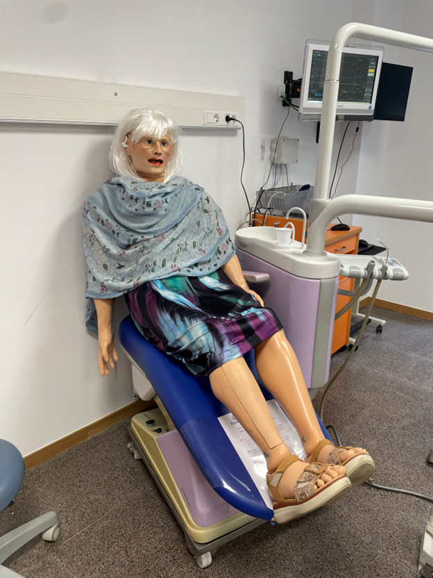
High-fidelity adult simulators used in dental education.The SimMan Essential simulator (Laerdal, Stavanger, Norway)
Inclusion and exclusion criteria
The inclusion criteria were: students enrolled in the subject Special Needs Dentistry and Gerodontology during the academic year and students who gave their authorization to be recorded for educational purposes. The exclusion criteria were students enrolled in only one of the subjects (Special Needs Dentistry or Gerodontology), and students who had already completed either of the two subjects, students who did not give their consent to be recorded for educational purposes. The activities carried out were assessed as part of the course, but only those students who agreed to participate in the study were included in the research.
Methodology
A total of six different clinical simulation scenarios were designed and implemented, each focusing on the management of medically compromised patients in a dental setting. Of these six scenarios, four involved the occurrence of a medical emergency during the dental appointment, requiring the students to respond promptly and appropriately within the clinical environment. The characteristics of each scenario—including the clinical focus, learning outcomes, simulation modality, and whether a simulated companion was present—are detailed in Table 1.
Table 1 Summary of clinical simulation scenarios
All students, in teams of 2–3, participated randomly in 1 or 2 simulation scenarios as part of the Special Care Patients course, and six months later, these scenarios were repeated and re-evaluated during the Gerodontology course, which shares content and instructors.
The methodology followed for each of the simulation scenarios was as follows:
Prebriefing
Before the simulation day, a prebriefing session is conducted to introduce students to the principles of clinical simulation and prepare them emotionally and cognitively for the activity. This phase is essential for establishing psychological safety, clarifying expectations, and fostering a respectful and collaborative environment. Instructors explain the structure, objectives, and educational value of simulation-based learning, as well as the roles that students will assume during the sessions.During this session, students are also organized into subgroups and each team is assigned one of the clinical scenarios. They receive a scenario checklist that serves as the basis for preparing their scientific report. Each subgroup is responsible for researching the assigned clinical context and developing a scientific evidence report. While there is no page limit, the document must include up-to-date references, with each team member contributing at least two properly cited sources [18, 19].
Safe environment activity
Each simulation session begins with a safe environment activity in which both instructors and students participate to promote trust, emotional safety, and group cohesion. These activities may include icebreaker games such as “Simon says,” or inviting students to share light personal facts, like what they enjoy for breakfast. The aim is to reduce anxiety, enhance communication, and create a supportive space for experiential learning [19, 20].
Briefing
Immediately before the simulation scenario begins, a briefing is conducted. Students who have prepared the scientific evidence present a concise summary of the clinical case to the rest of the group. This includes a review of the patient’s background, relevant clinical details, and anticipated challenges. The goal of the briefing is to ensure that all participants understand the scenario’s context, align on objectives, and are mentally prepared to engage in the simulation with clarity and confidence [21, 22].
Before entering the simulation room, students were informed that they would be attending to a patient returning for a second dental visit. The initial clinical history, dental examination, radiographs, and interconsultations had already been completed during the first visit. Although students had not seen this information beforehand, they were expected to simulate requesting it, and it would then appear on the monitor inside the simulation room. Their main task was to review the available data and prioritize the most appropriate dental treatment to initiate at this second appointment. Some clinical details were intentionally withheld so that students would actively ask the simulated patient and/or accompanying person, encouraging realistic communication and clinical reasoning—just as in a real consultation [21].
Simulation Scenario
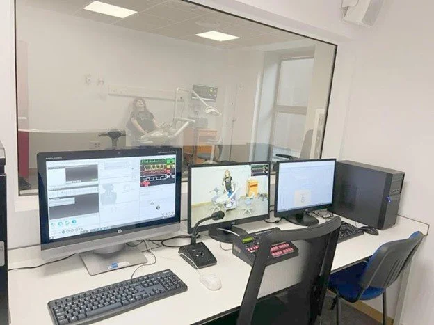
A team of 2 or 3 students enters the simulation room equipped with a dental chair and all necessary instruments to replicate a real clinical setting. The room is connected to the control room through a one-way mirror, allowing instructors to observe the simulation in real time without interfering with the scenario (Fig. 2), while the rest of the group remains in the debriefing room, where they can observe the entire simulation in real time through a video feed. Within the simulation team, roles are distributed to reflect a real clinical setting: one student acts as the dentist, another as the dental assistant, and, when applicable, a third student as the circulating assistant or second auxiliary.
Fig. 2
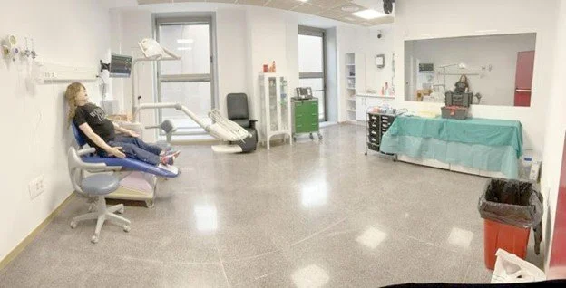
Dental simulation room connected to the control room through a one-way mirror
Before the simulation scenario begins, the student team is given approximately five minutes to familiarize themselves with the environment, review the available equipment, and organize the materials they may need. Then, the scenarios typically last between 10 and 15 min.
In the four scenarios that included a medical emergency, the critical event was programmed to occur at a specific point during the students’ clinical interaction—usually after reviewing the patient’s history or initiating treatment. The emergency (e.g., hypertensive crisis, hypoglycemia, asthma attack, epileptic seizure) was simulated through changes in the high-fidelity mannequin’s vital signs (e.g., blood pressure drop, respiratory distress, seizure activity) or verbal cues provided by standardized patients or instructors from the control room. Students were expected to recognize early warning signs, communicate effectively, and implement appropriate clinical protocols (e.g., positioning, oxygen administration, use of emergency kit, activation of emergency services). Instructors adjusted the scenario dynamically based on the students’ actions to maintain realism and ensure educational objectives were met.
Throughout the simulation, instructors are located in the control room (Fig. 3), where they manage the physiological parameters of the simulator and/or standardized patient, provide verbal responses when necessary, and observe student performance.
Fig. 3
Control room where instructors monitor and manage the simulation in real time
Evaluation is carried out in real time using standardized checklists designed by the research team for each simulation scenario. These checklists included both technical and non-technical competencies such as decision-making, teamwork, communication, and prioritization of care. The content of the assessment checklists was validated by an expert panel composed of 11 faculty members from six Spanish universities, all of whom are professors of the Special Care Dentistry course in their respective institutions, with a minimum of 10 years of clinical and teaching experience in this field. The experts independently reviewed each item using Likert-type scales to assess its importance and relevance for the evaluation of the scenarios. The checklists were then refined through a consensus process and piloted in prior simulation sessions before formal use in the study.
Debriefing
After the scenario ends, the participating students and the instructors return to the debriefing room. First, the students who performed the simulation share with the group how they felt, and then a guided and collective discussion begins to evaluate and reflect on the positive aspects and areas for improvement in the scenario they experienced.
In addition to the simulation itself, the methodology incorporated structured group learning and peer feedback components. During each session, while one team performed the simulation, the remaining students observed the scenario in real-time from the debriefing room. These students were encouraged to identify and record both effective actions and areas needing improvement. This process fostered collaborative reflection and discussion during the debriefing phase, where students collectively evaluated the scenario and exchanged perspectives. This approach enabled not only experiential learning but also peer-supported development of clinical and communication skills.
Evaluation
Evaluation of the students was carried out using the previously described assessment checklists. These checklists were completed twice: the first time during the subject Special Needs Dentistry, and a second time six months later, during the repetition of the clinical scenarios in the subject Gerodontology. All scenarios were recorded with the aim of using the recordings, if necessary, to improve the accuracy of the evaluation and during the debriefing session.
After collecting all evaluations from the three instructors, the results were compiled into Excel spreadsheets—one for each clinical scenario. The data were collected item by item, recording the evaluation provided (Yes/No) by the three evaluators in the initial simulation (PRE) and six months later (POST).
Data analysis
All the initial and final evaluation results of the students, obtained through the assessment checklists, were sent to the Statistical Support Section (SAE) of the University of Murcia. Data analysis was performed using R version 4.0.3. Normality and homoscedasticity (equality of variances) between the scores given by each observer in the initial and final evaluation of each assessment checklist were analyzed. For this purpose, Bartlett’s test and one-way ANOVA were used. Additionally, an average score per student was calculated based on the evaluations given by the three assessors for each checklist in both the initial and final assessments, and the p-value was calculated to determine the presence or absence of normality. Subsequently, either a paired t-test or a non-parametric paired test (if normality was not present) was used to determine whether the results were statistically significant.
Results
All students gave their consent to participate, resulting in a total of 64 participants included in the study. It is important to note that the assessments were conducted six months apart, during which time the students participated in clinical practice with real patients.
Evaluation checklist 1: hypertensive patient with hearing loss, hypertensive crisis
Table 2 displays the inter-rater agreement coefficients, p-values for each evaluator at both time points, and the result of Bartlett’s test. The initial scores did not follow a normal distribution, although homoscedasticity was confirmed. In contrast, the final scores met the assumptions of both normality and homoscedasticity. One-way ANOVA indicated no statistically significant differences among evaluators at either assessment point.
Table 2 Concordance coefficient for each evaluator for evaluation checklist 1 in the initial and final assessments, along with the results of bartlett’s test and significance levels
Fifteen students completed this scenario. Table 3 shows the mean scores of each student, calculated from the three evaluators’ ratings at both the initial and final time points. Most students showed improved performance in the final evaluation. A paired t-test revealed a statistically significant difference between the initial and final scores (p < 0.001).
Table 3 Average scores per student in evaluation checklists 1–6 across the two assessment phases
Evaluation checklist 2: patient with chronic bronchial disease, asthmatic crisis
The inter-rater agreement coefficients, the p-values for each evaluator, and the Bartlett test results are shown in Table 2. The initial scores followed a normal distribution, and the assumption of homoscedasticity was also met. However, the final scores did not meet the normality assumption, although homoscedasticity was preserved. No statistically significant differences were found between evaluators using the one-way ANOVA test.
In Table 3, the average scores obtained by the 17 students who completed Scenario 2 are shown. All scores, both individual and overall, were higher in the final assessment. Due to the lack of normality, a non-parametric paired test was applied, revealing statistically significant differences with a p-value of 0.003.
Evaluation checklist 3: patient with chronic renal failure
In Table 2, the inter-rater concordance coefficients and their significance levels for Evaluation Checklist 3 (patient with chronic renal failure) are presented. Bartlett’s test showed that the initial scores lacked normality but met the assumption of homoscedasticity. Similarly, the final scores also lacked normality, but homoscedasticity was maintained.
The one-way ANOVA revealed no statistically significant differences between evaluators at any stage of the assessment (Table 2).
Table 3 shows the average score obtained by each student in Scenario 3. All students demonstrated higher average scores in the final phase—during the repetition of the clinical scenario six months later—except for student number 21.
As normality was confirmed, a paired t-test was conducted, revealing statistically significant differences with a p-value < 0.001 (Table 2).
Evaluation checklist 4: diabetic patient, hypoglycemia
In Table 2, the concordance coefficients of the evaluators and their significance levels for Evaluation Checklist 4 (diabetic patient experiencing hypoglycemia) are shown. Bartlett’s test indicated that the initial scores did not meet the normality assumption, although homoscedasticity was confirmed. The final scores also failed to meet the normality assumption, but homoscedasticity was again confirmed.
Table 3 presents the average scores obtained by each student in Scenario 4. All average scores were higher in the final phase, corresponding to the repetition of the clinical scenario involving the diabetic patient six months later.
Given the lack of normality, a non-parametric paired test was conducted, yielding a p-value < 0.001. Therefore, the difference between the initial and final scores was statistically significant (Table 2).
Evaluation checklist 5: epileptic and anticoagulated patient, epileptic seizure
In Table 2, the concordance coefficients of the evaluators and their significance levels for Evaluation Checklist 5 (epileptic and anticoagulated patient experiencing an epileptic seizure) are shown. Bartlett’s test indicated that the initial scores did not meet the normality assumption, although homoscedasticity was confirmed. In contrast, the final scores met both the normality and homoscedasticity assumptions.
Table 3 presents the average scores obtained by each student in Scenario 5. All average scores were higher in the final phase, corresponding to the repetition of the clinical scenario involving the epileptic patient six months later.
A paired t-test was performed, yielding a p-value < 0.001, indicating that the results are statistically significant.
Evaluation checklist 6: patient with cerebral palsy and vision loss
In Table 2, the concordance coefficients of the evaluators and their significance levels for Evaluation Checklist 6 (patient with cerebral palsy and vision loss) are shown. Bartlett’s test revealed that the initial scores did not meet the normality assumption, although homoscedasticity was confirmed. In contrast, the final scores fulfilled both the normality and homoscedasticity assumptions.
Table 3 presents the average scores obtained by each student in Scenario 6. All average scores were higher in the final phase, corresponding to the repetition of the clinical scenario involving the patient with cerebral palsy and vision loss six months later.
Due to the lack of normality, a non-parametric paired test was applied. The results were statistically significant, with a p-value < 0.001.
Overall, across all six clinical scenarios, students demonstrated improved performance in the final assessments conducted six months after the initial evaluations. In each case, statistical analyses confirmed that the differences between initial and final scores were significant, regardless of whether parametric or non-parametric tests were applied. These results suggest a positive impact of the simulation-based training on students’ clinical competencies over time.
Discussion
Clinical simulation is being used in Health Sciences for various reasons, all focused on improving the quality of education and patient safety [23, 24]. In the Dentistry degree, low-fidelity simulators such as real teeth or oral cavity phantoms have been used for many years to practice clinical procedures (e.g., cavity preparation, fillings, endodontics) before performing them in the patient’s mouth [25, 26]. However, numerous authors argue that this type of simulation does not adequately allow for the development and evaluation of clinical performance, which encompasses both technical and non-technical skills, including teamwork, decision-making, communication, and leadership [26, 27].
In this study, four clinical scenarios (Scenarios 1, 2, 4 and 5) involving emergencies in the dental clinic were developed using the Laerdal SimMan Essential simulator (Laerdal, Stavanger, Norway). This simulator is an effective tool for improving students’ skills, knowledge, and attitudes, while also allowing instructors to assess clinical performance [1, 25, 27].
Additionally, standardized patients were used in Scenario 4 (diabetic patient), Scenario 5 (epileptic patient), and Scenario 6 (patient with cerebral palsy), since, as supported by Alinier [28], we believe that the inclusion of standardized patients provides an added dimension of realism and enables students to develop essential communication skills for clinical practice.
Numerous studies and theses have been found on the use of high-fidelity simulators and standardized patients in the training of students and professionals in Medicine and Nursing degree programs [9, 12, 29]. However, fewer studies specifically address their use in the Dentistry degree [1, 24, 25, 30].
Any dentist may face a dental emergency during their workday, and in such critical situations, coordination among the dental team is crucial to ensure the highest chance of patient survival. Dental emergencies are not very frequent, which makes high-fidelity clinical simulation an excellent method for training in these scenarios.
Roy et al. [1] implemented a methodology similar to this study. Students were divided into groups of three pairs, with each pair participating in two random 20-minute scenarios, while the rest observed from an adjacent room. Three months later, the simulation group and a control group (42 fifth-year Dentistry students who had not participated in the initial simulation) underwent a new simulation test and were evaluated using assessment checklist, followed by a comparison of the scores. In this study, no control group was used. All students, in teams of 2–3, participated randomly in 1 or 2 simulation scenarios as part of the Special Needs Dentistry course, and six months later, these scenarios were repeated and re-evaluated during the Gerodontology course, which shares content and instructors.
Given the positive results obtained in the study, future research could explore the implementation of high-fidelity clinical simulation in other areas of the Dentistry curriculum, such as pediatric dentistry. In this context, simulation could help students practice managing pediatric patients with behavioral issues or learn how to appropriately interact with the children’s family members.
The study has both limitations and strengths. Among its strengths is the longitudinal design, which evaluates students at two different time points six months apart, providing valuable information on long-term knowledge retention. Additionally, the combination of high-fidelity simulation and standardized patients allowed for the development of both technical and non-technical skills. Finally, the use of three independent evaluators and validated checklists ensured an objective and reliable assessment of student performance. A limitation of the study was the lack of an option to grade students with a “partially completed” item, often requiring a choice between marking it as done or not done, even when the task had been partially completed or there was an attempt to complete it, but the execution was inadequate. Perhaps if such an option had been available, the scores might have differed. Additionally, students participated in clinical practice with real patients during the six-month interval between the initial and final simulations. This clinical exposure may have contributed to their improved performance in the second round of simulations, making it difficult to isolate the specific effect of simulation alone.
Conclusion
Based on the results obtained in this study, which evaluated the effectiveness of clinical simulation as a learning tool in the Special Needs Dentistry course within the Dentistry degree program, the following conclusions can be drawn:
High-fidelity clinical simulation helps improve students’ clinical performance over time, particularly in applying theoretical knowledge effectively in simulated practice.The average scores obtained by students were higher in the final phase (six months later) compared to the initial phase.
In all scenarios, there was adequate concordance among evaluators, with no statistically significant differences, supporting the reliability of the evaluation process used.
Overall, we can conclude that simulated clinical scenarios, when used as a repetition tool in Dentistry, represent an effective pedagogical strategy, particularly highlighting their value in key areas such as dental emergencies or the treatment of patients with special care needs.
Data availability
No datasets were generated or analysed during the current study.
References
1.Roy E, Quinsat VE, Bazin O, Lesclous P, Lejus-Bourdeau C. High-fidelity simulation in training dental stduc. 2018;22(2):e261–8.
2. Kishimoto N, Sanuki T, Liu Y, Tran SD, Seo K. Simulation training for medical emergencies of dental patients: a review of the dental literature. Jpn Dent Sci Rev. 2023;59:104–13.
3. Kim J, Park JH, Shin S. Effectiveness of simulation-based nursing education depending on fidelity: a meta-analysis. BMC Med Educ. 2016;16:152.
4. Cleland JA, Abe K, Rethans JJ. The use of simulated patients in medical ed.